Nature Methods’ method of the year 2023, SenzaGen, groundbreaking “body-on-chip” by the University of Edinburgh and more
News on non-animal methods
Jan 2-5, 20241. Nature Methods’ method of the year 2023 : In vitro approach for modelling development
How a new organism can develop and what goes wrong in the context of congenital disorders has fascinated men for centuries. Development is a highly complex process that requires the precise interplay between various entities to ultimately form a proper embryo consisting of various cell and tissue types. One main challenge in the study of embryonic development is the access to suitable model systems.
Over the last years, in vitro models of embryonic development, or embryo-like structures, have been established that each recapitulate certain stages of embryonic development. These model systems are generated from stem cells : unspecified cells that can be maintained in the lab for a long time. These in vitro models have several advantages. For this reason, Nature Methods Journal has selected “methods for modelling development” as “Method of the Year 2023”. These methods clearly have a major impact on the scientific community, and also on society in terms of the potential to develop medical interventions in the future.
2. SenzaGen secures SEK 1.7m order from a new global biotech industry leader
SenzaGen has been selected to test substances for yet another new global customer within the biotech industry. The assignment is valued at SEK 1.7 million and includes non-animal tests for skin sensitization, utilizing SenzaGen’s highly performing test platform, GARD®skin. The tests will be conducted at the company’s GLP-certified laboratories in Lund and at the subsidiary, VitroScreen, in Milan during the fourth quarter of 2023 and the first quarter of 2024.
3. 3D-printed chip showing body’s reaction to drugs could end need for animal tests
Thousands of animals are used in the early stages of developing medicines worldwide every year, yet many drugs tested on animals do not end up showing any clinical benefit.
Now researchers at the University of Edinburgh have designed a groundbreaking “body-on-chip” that perfectly mimics how a medicine flows through a patient’s body. The device invented in Edinburgh is the first of its kind in the world.
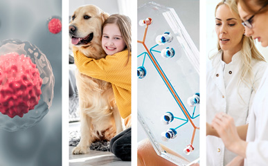
The plastic device means scientists can test drugs to see how different organs react without the need for live animal testing. Made using a 3D printer, the chip’s five compartments replicate the human heart, lungs, kidney, liver and brain. The plastic device uses positron emission tomography (PET) scanning to produce detailed 3D images showing what is going on inside the tiny organs.
4. Wyss Institute : Using AI, researchers identify a new class of antibiotic candidates
Using a type of artificial intelligence known as deep learning, researchers at the Wyss Institute and MIT have discovered a class of compounds that can kill a drug-resistant bacterium that causes more than 10,000 deaths in the United States every year.
In a study appearing today in Nature, the researchers showed that these compounds could kill methicillin-resistant Staphylococcus aureus (MRSA) grown in a lab dish and in two mouse models of MRSA infection. The compounds also show very low toxicity against human cells, making them particularly good drug candidates.
A key innovation of the new study is that the researchers were also able to figure out what kinds of information the deep-learning model was using to make its antibiotic potency predictions. This knowledge could help researchers to design additional drugs that might work even better than the ones identified by the model.
5. UPM Biomedicals enters into distribution agreement with Brinter for GrowInk bioinks
UPM Biomedicals, the forerunner in producing high quality nanofibrillar cellulose for life science and clinical applications, has signed an extensive distribution agreement for GrowInk™ bioinks with Brinter Inc, a Finnish-US medtech/biotech company focusing on the development of cartilage implants using their patented 3D bioprinting technology.
The agreement grants Brinter Inc the distribution rights of GrowInk bioinks in more than 30 countries including the United States, Canada, Finland, Austria, Belgium, Bulgaria, Denmark, Estonia, France, Germany, Netherlands, Sweden, Australia, New Zealand…
Advances in 3D bioprinting have been outstanding in the past decade. The technology is becoming widely used for various applications, and 3D bioprinting is already important in areas such as cancer research, where tumour models can be printed to test their response to different treatments.
6. IBEC to Develop Organs-on-a-Chip in Three Pathfinder Projects
BuonMarrow, OMICSENS, and PHOENIX-OoC are the three projects in which IBEC’s Biosensors for Bioengineering Group will apply its extensive knowledge in the field of biosensors and organs-on-a-chip. The projects, which will be developed with funding from the European Innovation Council’s prestigious Pathfinder Open program, promise to enhance cancer treatments and foster innovation in diagnostics.
While each project has specific goals, they all leverage advanced technologies such as biosensors, microfluidics, and artificial intelligence. These technologies not only represent significant scientific advances but also have the potential to directly impact patients’ lives by improving the efficacy of treatments, enabling earlier diagnoses, and facilitating the development of more accurate therapies.
7. Wyss Institute : A malaria drug treatment developed with Human organ on a chip could save babies’ lives
To try to reduce the risk of malarial infection, the World Health Organization recommends that pregnant women in low-income countries be treated with a combination of the antimalarial drugs sulfadoxine and pyrimethamine (SP). Curiously, a recent study found that this treatment also seemed to increase the birth weight of treated mothers’ babies, regardless of whether they contracted malaria.
Intrigued by this finding, with support from the Bill and Melinda Gates Foundation, a team of scientists at the Wyss Institute decided to investigate the phenomenon using its human Intestine Chip.
The Intestine Chip was created by taking healthy intestinal cells donated by female patients and culturing them inside a human Organ Chip device. This process allowed the team to create the first in vitro model of the adult female intestine and replicate the hallmarks of malnutrition to find treatments.
Human organ chip research shows that a common antimalarial combination could reverse the negative effects of malnutrition in the female digestive tract that lead to low birth weight infants. The research is published in eBioMedicine.
8. University of Toronto researchers target mutation underlying cardiac muscle disease using heart-on-a-chip
A team including researchers across the University of Toronto has developed a heart-on-a-chip device to study the effects of a genetic mutation that causes dilated cardiomyopathy, a heart muscle disease that impairs blood flow throughout the body.
Until now, researchers have had limited capacity to study cardiac muscle cells due to incomplete tissue maturation in the lab. The U of T‑led research team was able to mature stem cells into adult cardiac tissue to observe the effects of a sodium channel mutation that disrupts regular electrical activity in the heart.


