EU Roadmap : Key Recommendations, First Roadmap on OoC Devices, Awards to Non-Animal Methods, Artificial Organs Closer to Reality and more
News on non-animal methods
NEWS, REPORTS & POSITION STATEMENTS
1. Accelerating the transition : Key recommendations the EU must adopt
The EU’s approach to phasing out animal testing needs rapid acceleration, and by doing so the EU can set a new industry standard in chemical safety assessments. With the development and adoption of New Approach Methodologies, animal testing no longer needs to be seen as the “gold standard”.
In this new Frontiers Policy Labs commentary, Love Hansell, J. Ritskes-Hoitinga, I.J. Visseren-Hamakers, and Thomas Hartung, explore the key recommendations the EU must adopt in order to accelerate the transition to a world of chemical safety assessments void of animal testing, ultimately making them more human-relevant, efficient, and cruelty-free.
Read more in Frontiers Policy Labs
2. The first roadmap on organ-on-chip devices
For 2 years and with the support of NEN, more than 120 European experts worked on establishing the first Roadmap on organ-on-chip devices. Organ-on-Chip (OoC) aims to develop high-tech solutions mimicking the function of an organ using microchip technology.
Standardization is key for the further development of this field. For this purpose, the Focus Group Organ-on-Chip (FG OoC) has advised submitting the work program for adoption to the International Organization for Standardization (ISO). By choosing standardization at the international level, stakeholders recognize the broad global interest in the field and the various initiatives already underway. This acknowledges the global value chain for OoC technologies, and ensures that standards are developed with input from stakeholders worldwide, thus promoting innovation, interoperability, and safety.
Read the report and download the roadmap
3. NIH-Funded Centers Look to Make Tissue Chips FDA-Ready Tools
NCATS (National Center for Advancing Translational Sciences) has established new centers to strengthen the use of organ-on-a-chip, or tissue chip, technology to develop drugs and potentially reduce the need for animals in research. NIH has awarded approximately $31 million over five years to support four Translational Centers for Microphysiological Systems (TraCe MPS) at the University of Washington, Seattle and the Icahn School of Medicine at Mount Sinai in New York ; the University of Rochester ; the University of Pittsburgh ; and Texas A&M University, College Station and The University of Texas Medical Branch (UTMB), Galveston.
The TraCe MPS program is a partnership with the U.S. FDA. The FDA will work with each funded group to help it meet certain requirements for the chips to qualify as FDA-sanctioned drug development tools. “The centers are the last step in moving tissue chips into widespread use,” said Danilo Tagle, Ph.D., director of the NCATS Office of Special Initiatives. “The use of animal models hasn’t had an especially good success rate in developing drugs. Currently, tissue chips are mostly considered complementary to the use of animals in research. This has already begun to change.”
INTERVIEWS, NOMINATIONS & AWARDS
4. ERC Proof of Concept grant to develop a stem cell-based screening assay to study male fertility
Prof. Susana Chuva de Sousa Lopes, reNEW Leiden PI, receives an ERC Proof of Concept grant that allows her to develop a stem cell-based screening assay for toxic effects of chemical compounds and environmental factors on male fertility. This will provide a valuable alternative to current expensive, time-consuming and often unreliable toxicity testing in animal models.
The assay – ReproTox – leverages the differentiation of hiPSCs to sperm progenitor cells which are subsequently exposed to chemicals and environmental factors to assess their effect on male fertility. The assay can in the future be used by the private sector to assess toxicity during early stages of drug development and thereby provides financial, ethical and societal benefits.
5. Non-animal method with regulatory approval for evaluating hormone potency awarded 3Rs Prize
This year’s UK 3Rs Prize has been awarded to Dr Francesco Nevelli at Merck for a publication describing the development of an in vitro assay for testing follicle-stimulating hormone product potency. The in vitro method was approved by regulators for use in potency testing, at best replacing tens of thousands of animals annually.
Francesco and colleagues are now working towards worldwide acceptance to enable Merck to fully replace all rats used for potency tests of Merck Healthcare marketed products, with estimated saving of nearly 40,000 animals annually, based on forecast animal-use in 2023. The Prize money will be used to fund educational programmes for physicians and patients in the USA to increase acceptance of in vitro quality control processes for biological products.
Read more on the awarded programs
TOOLS, PLATFORMS, CALLS
6. 2025 Descroix-Vernier EthicScience Prize : extended deadline
Created in 2013 by the Pro Anima Scientific Committee, the EthicScience Prize became the Descroix-Vernier EthicScience (DVES) Prize in 2023, marking the convergence of the objectives and values of Pro Anima and the Fondation Descroix-Vernier.
With a total endowment of 110,000 euros rewarding 3 categories (Innovation ; Development and Applicability ; Jury’s Prize — Forward-Looking Project), the DVES prize is one of the best endowed prizes in Europe entirely dedicated to the most advanced, most convincing scientific and technological knowledge for alternatives to animal experiments. A multidisciplinary and multi-sector strategic committee, chaired by Dr Jean-Pierre Cravedi (Toxicologist, Chairman of Aprifel’s Scientific Advisory Board, former expert at ANSES and EFSA) ensures the quality, transparency and impact of the Prize.
The DVES 2025 Prize is still open for application with an extended deadline : September 30, 2024.
7. Advances in animal-free models and data analysis for drug development
The ever changing landscape of animal-free research is increasingly gaining momentum and relevance by the recent announcement of the FDA that some animal tests before human drug trials may no longer be required.
In light of these developments, a special edition invites submissions covering reviews on advances of animal-free models and data analysis for drug development focussing on human diseases and regeneration. This includes in vitro/ex vivo modelling, bioprinting, the use of 3D structures such as spheroids, organoids or cellular scaffolds, software development, mathematical modelling and computational approaches that support animal-free, alternative drug development.
Deadline : 31st January 2025
8. Lab of the Future Survey 2024
If you’re a member of the Pistoia Alliance or plan to be, take the opportunity to contribute to its annual global Lab of the Future survey 2024 about the adoption of emerging technologies and the skills that will be needed to enable innovation and collaboration within the labs of the future.
The Lab of the Future survey, below, takes 6 minutes to complete. All survey participants will receive a copy of the published report.
9. WORC.Community : The network for organoid and organ-on-a-chip researchers
In a rapidly advancing and transformative area of science, WORC.Community (World Organoid & Organ-on-chip Research Community) has been developed to enable communication and strong networks as essential parts to make informed decisions and discoveries. WORC.Community works to advance medical science, reducing and replacing the animals used in research and in supporting the innovative researchers and technologies.
INDUSTRY, BIOTECH & PARTNERSHIPS
10 . InSphero, Breakthrough T1D and Université Libre de Bruxelles : New cures for type 1 diabetes
InSphero, a leading innovator in 3D Cell Culture and Organ-on-Chip technology, has announced a new collaboration with Breakthrough T1D, the leading global type 1 diabetes (T1D) research and advocacy organization, and the Université Libre de Bruxelles (ULB) Center for Diabetes Research. With funding from Breakthrough T1D under its Industry Discovery & Development Partnerships (IDDP) program, this collaboration aims to uncover novel treatment strategies for T1D, focusing on protecting and preserving the vital insulin-producing beta cells in the pancreas.
11. Biomatter raises €6.5M to revolutionize enzyme design with generative AI
Biomatter, a pioneer in enzyme design, has successfully raised €6.5 million in a seed funding round co-led by Inventure and UVC Partners. This milestone marks a significant leap in the application of generative AI in synthetic biology, setting the stage for groundbreaking advancements in the field.
“Enzymes will play a paramount part in the future of the bioeconomy — they are the enabling piece that ultimately allows us to create new molecules, cells, and organisms for the world. The unique enzymes we have successfully developed for our global partners so far demonstrate our capability to go far beyond the existing enzyme improvement paradigm. At Biomatter, we believe that the unlimited capacity to design new enzymes will help to build a better tomorrow for everyone” said Laurynas Karpus, Co-founder and CEO of Biomatter.
SCIENTIFIC DISCOVERIES & PROTOCOLS
12. 3‑D reconstructed tissue model to assess the irritation and phototoxicity
This study introduces a novel in vitro methodology that employs the 3‑D reconstructed tissue model, EpiOcular, to assess the irritation and phototoxicity potential of medical devices and drugs in contact with the eye. The study evaluated diverse test materials, including medical devices, ophthalmological solutions and an experimental drug (cemtirestat), for their potential to cause eye irritation and phototoxicity.
By developing and evaluating these protocols for medical devices, the authors aimed to expand the applicability domain of the tests referred to in ISO 10993 – 23. This will contribute to the standardisation and cost-effective safety evaluation of ophthalmic products, while reducing reliance on animal testing in this field. The findings obtained from the EpiOcular model in the photo-irritation test could support its implementation in the testing strategies outlined in OECD TG 498.
13. Consensus-driven systematic review by the ProEuroDILI network
Idiosyncratic drug-induced liver injury (DILI) is a complex and unpredictable event caused by drugs, and herbal or dietary supplements. Early identification of human hepatotoxicity at preclinical stages remains a major challenge, in which the selection of validated in vitro systems and test drugs has a significant impact.
In this systematic review, the authors analyzed the compounds used in hepatotoxicity assays and established a list of DILI-positive and ‑negative control drugs for validation of in vitro models of DILI, supported by literature and clinical evidence and endorsed by an expert committee from the COST Action ProEuroDILI Network.
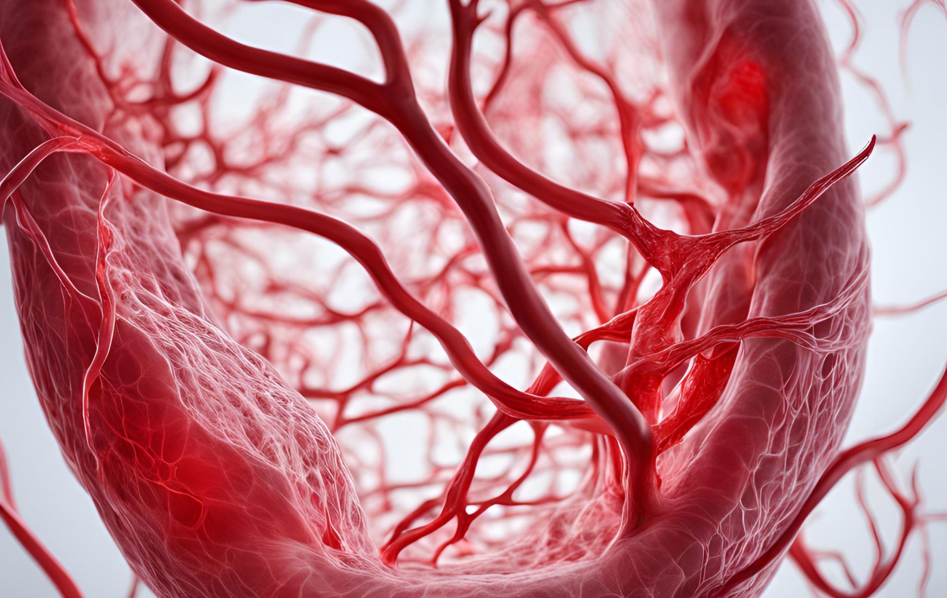
14. 3D-printed blood vessels bring artificial organs closer to reality
New research from Harvard’s Wyss Institute for Biologically Inspired Engineering and John A. Paulson School of Engineering and Applied Science (SEAS) brings that quest one big step closer to completion. A team of scientists created a new method to 3D print vascular networks that consist of interconnected blood vessels possessing a distinct “shell” of smooth muscle cells and endothelial cells surrounding a hollow “core” through which fluid can flow, embedded inside a human cardiac tissue.
“To say that engineering functional living human tissues in the lab is difficult is an understatement. I’m proud of the determination and creativity this team showed in proving that they could indeed build better blood vessels within living, beating human cardiac tissues. I look forward to their continued success on their quest to one day implant lab-grown tissue into patients,” said Wyss Founding Director Donald Ingber, M.D., Ph.D. Ingber.
Read the publication in Advanced Materials
15. Exciting advances in stem cell therapy
A new technique developed by McGill researchers for mechanically manipulating stem cells could lead to new stem cell treatments, which have yet to fulfill their therapeutic potential. “The great strength of stem cells is their ability to adapt to the body, replicate and transform themselves into other kinds of cells, whether these are brain cells, heart muscle cells, bone cells or other cell types,” explained Allen Ehrlicher, an associate professor in McGill’s Department of Bioengineering and the Canada Research Chair in Biological Mechanics. “But that is also one of the biggest challenges of working with them.”
Recently, a team of McGill researchers discovered that by stretching, bending and flattening the nuclei of stem cells to differing degrees, they could generate precisely targeted cells that they could direct to become either bone or fat cells.
Read the publication in Biophysical Journal
16. Cancer-on-chip : A 3D model for the study of the tumor microenvironment
The approval of anticancer therapeutic strategies is still slowed down by the lack of models able to faithfully reproduce in vivo cancer physiology. On one hand, the conventional in vitro models fail to recapitulate the organ and tissue structures, the fluid flows, and the mechanical stimuli characterizing the human body compartments. On the other hand, in vivo animal models cannot reproduce the typical human tumor microenvironment, essential to study cancer behavior and progression.
This study reviews the cancer-on-chips as one of the most promising tools to model and investigate the tumor microenvironment and metastasis. Pros and cons of this technology are also discussed highlighting the future challenges to close the gap between the pre-clinical and clinical studies and accelerate the approval of new anticancer therapies in humans.
Read the publication in Biological Engineering


